OUTLINE
Define
Avascular necrosis of the femoral head is a disease of the group of osteonecrotic diseases with the death of cells in both parts of the bone, the hematopoietic fat marrow and the bone cells.
The term osteonecrosis of the femoral head was first described by Munro (1738), then Cruveilhier (1835) described altered femoral head morphology secondary to disruption of blood circulation. Avascular necrosis of femoral head is also known as femoral head necrosis, ischemic necrosis, avascular necrosis, osteochondritis.

Epidemiology
Avascular necrosis of the femoral head is diagnosed at a rate of 10,000 to 20,000 new cases per year in the United States, with about 5–10% per year being avascular necrosis of the femoral head out of a population of 500,000. hip joint completely replaced in the US. In Asian countries, it is higher as in 2000, 36% out of 892 total hip replacements at National Taiwan Hospital were avascular necrosis of the hip. Bilateral femoral head necrosis accounts for 33-72%.
Risk factor
1. Directly: irradiation, trauma, blood diseases, pressure disorders – Caisson disease, marrow replacement disease (eg, Gaucher disease), sickle cell disease.
2. Indirect: alcohol abuse, hypercoagulable states, steroids (endogenous or exogenous), systemic lupus erythematosus, organ transplant recipients, viral infections (CMV, hepatitis, HIV, measles, chickenpox), inhibitors protease (a drug that treats HIV), spontaneous.
REASON
Gender and age
Avascular necrosis of the femoral head can occur at any age. However, the majority of cases occur in men, under the age of 50. The male-to-female ratio of this disease is about 8:1. The mean age of illness in women is about 10 years higher than in men.
Causes of the body's ability to supply oxygen
Lesions of this disease can occur in one hip or both sides. The disease accounts for about 10% of total hip replacements annually worldwide.
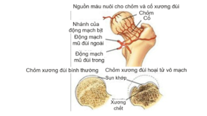
The femoral head and neck are supplied by the obturator artery and the circumflex artery. These are branches of the internal iliac artery and the deep femoral artery. Osteonecrosis occurs when there is insufficient oxygen supply in the diseased bone tissue.
Avascular necrosis of the femoral head usually occurs in the fatty marrow of the bone, where there are few blood vessels to nourish. The most vulnerable area is the anterior superior portion of the femoral head, just below the load-bearing surface of the femur.
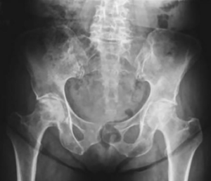
Injured bone shows death of the trabeculae and bone marrow, sometimes extending to the subchondral bone. The damaged bone is not able to completely restore the vascular system. When there is damage on X-ray There is usually a flattening of the crown that occurs after a few weeks to a few years.

Physiological or pathological causes
Necrotizing femoral head disease can be spontaneous or also be the result of some physiological or pathological conditions such as pregnancy, alcohol consumption, embolism, trauma, drug use, etc.
MECHANISM OF DISEASE
The pathogenesis of femoral head necrosis involves several mechanisms:
- Embolism or thrombosis in the small arteries of the femoral head. Caused by fat droplets, red blood cell adhesions, air bubbles.
- Destruction of vascular wall structure due to vasculitis, radiation necrosis, or release of vasoconstrictor factors.
- These injuries lead to a decrease or loss of blood supply to the bone tissue. Next, the body has a phenomenon of congestion in neighboring bone organizations leading to loss of minerals and a decrease in the volume of the bones. Bones are prone to collapse under pressure. This process takes place continuously leading to joint destruction after 3-5 years if not detected and treated.
CLINICAL
Necrotizing femoral head disease may progress without clinical manifestations. Bone lesions, detected on magnetic resonance imaging, may precede clinical manifestations by weeks to months.
Symptoms often appear in the advanced stages of the disease with pain or tenderness in the groin or around the affected hip. If the stage is late, the patient may have limited hip mobility and limp gait.
The physical symptoms are not specific. In the early stages, the patient may have limited range of motion of the injured hip. The patient has pain on examination, especially with rotation or hip flexion. Patients can maintain movement of the injured hip for many years with limited range of motion. As the disease progresses, patients often have severe pain, stiffness, or limited range of motion in the hip. The time to progression of the disease from onset to late stage varies from several months to several years.
SUBCLINICAL
To perform laboratory testing for this disease, you may need imaging tests or biochemical tests:
Imaging tests
1. Plain X-ray of the hip
Performing radiographs of the hip is often diagnostic in advanced stages of the disease. However, this method has little diagnostic value in the early stages of the disease. There are two X-ray positions of the hip that can help detect lesions: anteroposterior hip arthroplasty and lateral hip arthroplasty with frog leg position. Radiographs can often be normal many months after the appearance of the first clinical signs.
2. Bone scan
This method has diagnostic value for patients with suspected necrotizing femoral head lesions but normal X-ray images. Signs of bone scintigraphy specific for femoral head necrosis include:
- Image of hyperintensity due to new bone formation or increased metabolism around necrotic bone tissue.
- The image of "donuts" or images of cold spots in hot areas is less common, but extremely characteristic for femoral head necrosis.
3. CT-scan
CT scan can help detect avascular necrosis of the femoral head early. With fibrosis in the central region of the crest, the physician can assess the size of the necrotic area, detecting a slight collapse of the anterior part.
4. Magnetic Resonance Imaging
This is the most valuable method for detecting avascular necrosis of the femoral head. Patients can be performed at an early stage in the absence of a collapsed head and abnormal imaging tests. The sensitivity of this method is up to 90%.
Biochemical tests
The blood and biochemical tests in the patient with necrosis of the femoral head were completely normal.
STAGES OF Illness
The disease stage of femoral head necrosis is usually based on imaging studies and histopathology.
- According to Ficat and Arlet (1997) there are four degrees based on the radiographic appearance of the femoral head (in 1985, it was extended to stage 0).
+ Grade 0: detected based on biopsy only.
+ Grade 1: X-ray is normal, diagnosis is based on CT-scan, MRI.
+ Grade 2: Abnormal X-ray, no collapsed head.
- 2a: Distinctive skeletal features, with bright recesses.
- 2b: Sign fracture subchondral, showing a crescent-shaped light line.
+ Grade 3: Collapsed femoral head, subchondral bone fracture.
+ Degree 4: Osteoarthritis secondary, deformed femoral head, affecting the acetabulum.

- According to ARCO (Association Reseach Circulation Osseous) (1993) proposed division into 6 stages (or 6 degrees) and this classification system is currently the most commonly used.
- Stage 0: Laboratory tests and diagnosis were normal. Diagnosis of this stage is based on histopathology, the presence of necrosis on bone biopsies.
- State 1: Normal X-ray and CT-scan were normal. Patients with abnormalities on bone scan or MRI, bone biopsy showed signs of necrosis. The patient may have clinical manifestations.
- Phase 2: There is a clear dead bone in the femoral head on X-ray. These images may include bands of necrosis, calcium deposits, and cysts in the femoral head or neck. The femoral head remains spherical on both anterior and posterior radiographs and on radiographs and CT scans.
- Stage 3: The femoral head has mechanical changes. The bone below the cartilage with a crescent moon appears. The crest still retains its spherical shape.
- Stage 4: There is any evidence of femoral head collapse with signs of joint space stenosis.
- Stage 5: Any or all of the features described may be present in addition to joint space stenosis. The patient presented with degenerative joint manifestations secondary to mechanical changes in the femoral head. Manifestations of fibrosis, bone cysts in the acetabulum and possibly bone spurs.
- Stage 6: Severe destruction of the femoral head with severe degenerative manifestations.
DIAGNOSE
Implementing the quadrants
Definitive diagnosis is based on epidemiological factors, age and sex, combined with clinical symptoms and imaging tests.
Differential diagnosis
In patients with stages 1 and 2, definitive diagnosis is often difficult. With stage 1, doctors need to distinguish from all diseases with damage to bone, cartilage and synovial membrane. In stage 2, nonspecific erosive lesions on radiographs should be identified by bone scintigraphy and MRI.
In pregnant women, it is important to differentiate femoral head necrosis from pregnancy-induced transient osteoporosis. With stages 3 and 4, the radiographic findings are specific for avascular necrosis of the femoral head. However, it is difficult for patients in stages 5 and 6 to distinguish necrosis of the femoral head from other hip diseases because the late stages of these diseases have similar manifestations.
TREATMENT
Treatment of femoral head necrosis is one of the most controversial issues. The goal of treatment for this disease is to preserve the natural structure of the joint for as long as possible. And the extent of damage to the femoral head will determine the treatment method:
- Lesions less than 15% volume of the head, apply conservative treatment measures
- Injuries from 15 – 30% with cap volume, treated with decompression drilling or osteotomy
- Injuries over 30% of head volume, the treatment method is total hip replacement surgery
Four treatments are applied to patients with avascular necrosis of the femoral head including:
Conservative treatment
This method applies to stage 0, 1 and 2 patients. This stage can be treated with conservative treatment or decompression drilling. Conservative treatment includes:
- Patient resting, reducing hip load by using crutches
- Pain relief with non-steroidal anti-inflammatory drugs or pain relief alone
- Incorporate physical therapy methods to maintain muscle strength, avoid contractures, and use walking aids.
However, this measure does not help stop the progression of the disease.
Hip replacement surgery
Hip replacement surgery is applied to patients with persistent pain, poor response to treatment, and rapidly progressive loss of motor function.
This method should be performed before the femoral head is completely collapsed, usually at stages 3 and 4 (according to Ficat).
Top pressure relief drill
This measure is usually applied to patients in stages 1, 2 and 3 (according to ARCO) and patients who have applied conservative treatment but have not been effective.
The purpose of this measure is to reduce the pressure in the bones, rebuild the bone perfusion system. At the same time, this method creates conditions for the adjacent healthy bone to participate in the bone regeneration process.
The good results of this method account for 30 – 90% cases.
Bone shaping
This is a technique aimed at preserving joints. This method removes the diseased bone in the main load-bearing area of the femoral head in order to redistribute the pressure on the articular cartilage supported by the underlying healthy bone tissue.
Bone grafting, stem cells
This method is a new treatment that uses bone grafting techniques without vascular regeneration or stem cells. The aim of the method is to restore the necrotic bone organization of the head, from which the patient can Rehabilitation of the injured hip. However, this method is still in the experimental phase.

