Table of contents
ABSTRACT
Objective: find out the form and extent of damage meniscus due to endoscopic trauma knee joint.
Objects and methods: prospective study of 106 patients (patients), diagnosed meniscus tear due to trauma and laparoscopic surgery at Thong Nhat Hospital, Ho Chi Minh City since July 20067/2011.
Results: damage to the meniscus accounted for the majority with 72 cases (68%). The rate of tearing in the posterior horn of meniscus is the highest, 61/106 cases (accounting for 48.2%), the most common anatomical lesions are: longitudinal tear accounted for 55/106 cases (accounting for 51.9%). . Distribution of tear areas: the red-red area has 24/106 cases (accounting for 22.6%), the red-white area has 38/106 cases (accounting for 35.9%), the white-white area has 44/ 106 cases (accounting for 41.5).
Conclusion: damage to meniscus in trauma often damages the inner meniscus, especially the posterior horn, the common anatomical morphology is longitudinal tear and the red-red and red-white areas account for a high percentage.
Key word: Arthroscopy, meniscus damage.
ABSTRACT
STUDY ON THE TEAR MENISCUS IN THE KNEE TRAUMA WITH ARTHROSCOPY
Vo Thanh Toan, Nguyen Tien Binh, Tran Dinh Chien
* City Medicine. Ho Chi Minh * Vol. 16 – Supplement of No 1 – 2012: 226 – 230
Objective: to study the meniscus damage in the knee trauma with arthroscopy.
Methods: a prospective study was carried out on 106 patients who had diagnosed meniscus tear due to trauma and were arthroscopic surgery at Thong Nhat Hospital, Ho Chi Minh city, from july 2006 to july 2011.
Results: we had the results: radial meniscal tears with 72 cases (68%). The rate of meniscal posterior horn in the highest 61/106 cases (48.2%), the most common: vertical tearing 55/106 cases (51.9%). Distribution torn areas: the red-red: 24/106 cases (22.6%), the red-white: 38/106 cases (35.9%), the white-white: 44/106 cases (41.5%).
Conclusions: In Trauma, meniscus tears are often radial meniscal tears, vertical tearing. Distribution torn
* Ho Chi Minh City Thong Nhat Hospital ** Military Medical Academy
Author contact: MSc Vo Thanh Toan Tel: 0918554748 Email:
zone: the red-red and the red-white zone high percentage.
Keywords: Arthroscopy, Meniscus damage
QUESTION
The first arthroscopic knee surgery was performed in the world on March 9, 1955 by Watanabe M.(3). Since then, laparoscopic surgery has made many developments
quickly, gradually perfected and widely applied today thanks to many advantages: not only helps accurately diagnose injuries inside the knee joint, but also treats those injuries.
In our country, in recent years, along with the increase in means of transport and the growing and extensive movement of exercise and sports, the number of knee injuries in general and meniscus injuries has increased. in particular is increasing. Traumatic meniscal injuries are more common than other types of meniscus injuries, accounting for 68-75%(5).
Accurate diagnosis of meniscus damage and extent is very important because it helps surgeons make appropriate and timely diagnosis and treatment methods, which is of great significance in avoiding Unnecessary consequences arising from this injury such as limited movement of the knee joint, muscle atrophy, osteoarthritis as well as the rehabilitation of the knee joint.
At Thong Nhat Hospital, Ho Chi Minh City, since July 2006, arthroscopic knee surgery has been performed. Therefore, we carried out this study with the aim of understanding the morphology and extent of meniscal injury through knee arthroscopy.
AUDIENCE – METHODS
Research subjects
106 patients were diagnosed with meniscus tear due to trauma and laparoscopic surgery from July 2006 to July 2011 at Thong Nhat Hospital, Ho Chi Minh City.
Method of study
Study design: prospective study
Steps to take
Arthroscopic knee surgery
Necessary surgical tools: dipstick for inspection, scissors, pair of pliers, gnawing pliers with left, right or straight angles to treat meniscus lesions at different locations.
Anesthesia method and patient's position: The patient was given spinal anesthesia. The patient's position is supine, the thigh is firmly fixed to a support in the middle 1/3 contiguous to the lower 1/3 of the thigh. Garo is placed in the middle 1/3 of the thigh. The pillow can be folded up to 100°.
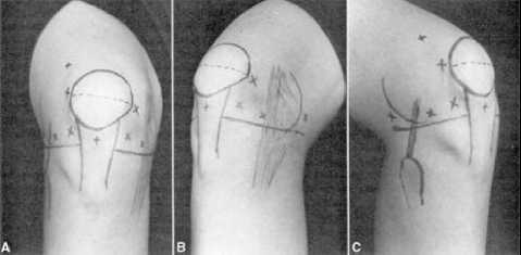
Technique: First of all, the patient lies on his back, the knee joint is in a 90 . flexion positiono, determine anatomical landmarks for skin incision, thereby putting the instrument system into observation, exploration, for diagnosis and treatment, we usually use two lines.

Anterior lateral line: landmarks along the lateral border of the patellar tendon, diagonally along the lateral condyle of the femur, and along the anterolateral border of the tibial plateau. These three lines form a triangle, in the area of this triangle is the possible position of the knee joint. Using a knife, make an incision about 0.5cm long through the skin and subcutaneous tissue, insert the troca into the joint socket, open the valve for washing solution (sterile physiological termite water) into the joint, suction and wash until the water is clear.
Anterior-inner line: the landmark opposite the inside of the joint slot, use a knife to make 0.5cm skin incision, insert the probe into the joint to check, determine the morphology of the meniscus damage in the joint, the damage to the anterior horn position, middle or posterior horns and associated lesions if any such as cruciate ligament, facet joint, cartilage surface of femoral condyle, tibial plateau, patella, etc.
Sau khi đã xác định vị trí đường vào khớp gối, phẫu thuật viên đưa ống soi vào để quan sát theo một trình tự nhất định từng vị trí của khớp gối như: túi bịt cơ tứ đầu đùi, mặt khớp lồi cầu bánh chè, mặt sụn sau xương bánh chè, ngăn khớp phía ngoài, ngăn khớp phía trong, sụn chêm trong và mâm chày, lồi cầu trong xương đùi, vị trí hố liên lồi cầu để đánh giá tình trạng dây chằng chéo, sụn chêm ngoài, lồi cầu và mâm chày ngoài, phẫu thuật viên khi quan sát hết sức cẩn thận, tránh bỏ sót thương tổn.
Research on meniscal injury caused by knee arthroscopy
There are many ways to classify meniscal lesions, but according to us, we choose the classification methods that contribute to the orientation of management during surgery.
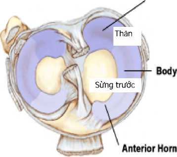
Studying the location of tearing according to the horns: anterior, posterior, and middle (body) horns.

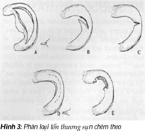
Research according to O'Connor's classification: considered the most complete(9), we apply this type of classification.
The tear line along the meniscus body: the tear line extends along the meniscus body, vertically, parallel to the meniscus border, the tear line can be the entire thickness of the meniscus or not.
Horizontal meniscal tear: The tear is horizontal, dividing the meniscus into two upper and lower parts.
Cross tear meniscal body: tear line of the entire thickness of meniscus, oblique diagonally from the inside to the body of the meniscus, can be diagonally anterior or posterior.
Fan-shaped tear line: vertical tear from the meniscus looks like a fan spoke, which can tear the full thickness of the meniscus or not at all.
Deformed tear line: tearing the entire meniscus, tearing with degeneration.

O'Connor(9). (A: longitudinal tear; B: cross tear; C: tearhorizontal; D: garbage h fan spokes; E: tear deformation). *Study the tear site according to the blood supply area of the meniscus:

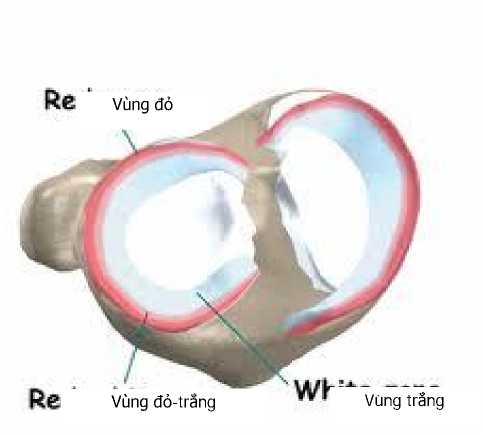
Area rich in blood vessels (red-red area): accounting for the outer third, this area is full of blood vessels, tearing this area is easy to recover if detected early and treated properly.
Medial area (red-white area): in the middle third of the blood vessel, the blood vessels begin to decrease gradually, the lesion can heal with the right treatment, but the results are lower.
Avascular area (white-white area): The inner third has no blood vessels, the tear here is not able to recover, so it is usually treated to remove the tear.
Data processing
Using SPSS 15.0 software.
RESULTS AND DISCUSSION
Damage to meniscus tear along the horn
Table 1: Damage to meniscus tear along the horn
|
Location |
Inner meniscus |
External meniscus |
Shared |
|||
|
SL |
TL% |
SL |
TL% |
SL |
TL% |
|
|
Front horn |
8 |
7,5 |
8 |
7,5 |
16 |
15 |
|
Middle horn (body) |
25 |
23,6 |
9 |
8,5 |
34 |
32,1 |
|
Rear horn |
37 |
35 |
14 |
13,2 |
61 |
48,2 |
|
Total |
2 |
1,9 |
3 |
2,8 |
5 |
4,7 |
|
Total |
72 |
68 |
34 |
32 |
106 |
100 |
We performed 106 knee arthroscopy (106 patients). Injury to the inner meniscus accounted for the majority with 72 cases (accounting for 68%) (table 1), this difference is statistically significant (p<0.05), this result is also consistent with the authors in this study. and abroad. This is also explained by the authors that: the posterior horn (16-20mm) is wider than the anterior horn (8-10mm), the anterior horn firmly adheres to the tibial plateau just in front of the anterior tibial spine and ligament. front cross. The posterior horn attaches to the posterior tibial plateau just anterior to the attachment of the ligament rear cross, closely related to lateral ligament posterior medial and semi-membranous tendons, etc. It is the anatomical relationship with the surrounding components that limits the movement of the medial meniscals (in contrast, the outer meniscus is more flexible) when flexing and extending the knee. This explains why damage to the medial meniscus is common in knee injuries(1,2,5).
Also according to Table 1, the rate of tearing in the posterior horn of meniscus is the highest in 61/106 cases (accounting for 48.2%), this difference is statistically significant (p<0.05), the results This is also suitable for some domestic authors such as: Nguyen Manh Khanh(7), Truong Kim Hung (11), Nguyen Quoc Dung(8) and foreign authors such as: Allaire(1), Tapper(10) also recognized that tearing of the posterior horn of meniscus, especially the lateral meniscus, is common, but the mechanism has not been clearly elucidated.
According to O'Connor's classification
Table 2: Classification of lesions
|
Injury form |
Inner meniscus |
External meniscus |
Shared |
|||
|
SL |
TL% |
SL |
TL% |
SL |
TL% |
|
|
Vertical tear |
37 |
34,9 |
18 |
17 |
55 |
51,9 |
|
Horizontal tear |
5 |
4,7 |
4 |
3,8 |
9 |
8,5 |
|
Cross tear |
20 |
18,9 |
6 |
5,7 |
26 |
24,5 |
|
Torn fan spokes |
3 |
2,8 |
1 |
1 |
4 |
3,8 |
|
Torn deformation |
7 |
6,6 |
5 |
4,7 |
12 |
11,3 |
|
Total |
72 |
67,9 |
34 |
32,1 |
106 |
100 |
According to Table 2, of the 106 knee joints we operated on, the most common anatomical injury was: longitudinal tear accounted for 55/106 cases (accounting for 51.9%) followed by cross tear: 26/106 cases (accounting for 51.9%) 24.5%) and the least common group is fan blade tear: 4/106 cases (accounting for 3.8%), this difference is statistically significant (p<0.05) consistent with the authors in this study. country and abroad, followed by cross-type: 26/106 cases (24.5%), which is slightly different from other authors. This can be explained: due to the different arrangement of collagen fiber bundles in different positions of the meniscus, it leads to different anatomical lesions: lesions at the meniscal margin. Close to the joint capsule is usually a longitudinal tear, the injury in the free part is usually a transverse tear or a flap tear. In addition, the type of meniscal injury depends on age as shown by the thickness and quality of the cartilage layer of the tibial and femoral facets. Young people have thick, elastic, and well-absorbed articular cartilage, so they often see longitudinal tears. Adults over 30 years old cartilage quality begins to decline, unable to absorb rotational forces, resulting in a horizontal tear or a diagonal tear. In the elderly, there is a lot of degeneration of the cartilage, the cartilage layer is lost, the joint space of the knee joint narrows, the rolling motion of the condyle on the tibial plateau is subject to much friction, so there is often a ragged tear.(5,4,6). In our study group, the group over 30 years old accounted for more than other authors, which could explain the second highest rate of our cross tear group after longitudinal tear group.
Distribution of tear area
Table 3: Distribution of tear area
|
Torn area |
SL |
TL% |
|
Red-red zone |
24 |
22,6 |
|
Red-white area |
38 |
35,9 |
|
White–white area |
44 |
41,5 |
|
Total |
106 |
100 |
Chúng tôi gặp cả 3 nhóm rách ở vùng đỏ- đỏ: 24/106 trường hợp (chiếm tỷ lệ 22,6%), vùng đỏ-trắng: 38/106 trường hợp (chiếm tỷ lệ 35,9%), vùng trắng-trắng: 44/106 trường hợp (chiếm tỷ lệ 41,5%), sự khác biệt này không có ý nghĩa thống kê. Kết quả này khác với một số tác giả trong nước (nhóm rách vùng trắng-trắng chiếm đa số), có thể do cơ chế gây tổn thương của nhóm BN chúng tôi nghiên cứu đa số là tai nạn giao thông, khác với các tác giả khác là do sports injury chiếm đa số.
CONCLUDE
Through 106 patients in the study, we draw some conclusions as follows:
Injury to meniscus tear along the horns: damage to the meniscus accounted for the majority with 72 cases (68%) of knee injuries. The rate of tearing in the posterior horn of meniscus is the highest: 61/106 cases (accounting for 48.2%)
Injury to torn meniscus according to O'Connor's classification: in the 106 knee joints we operated on, the most common type of anatomical injury was longitudinal tear, 55/106 cases (accounting for 51.9%).
Distribution of tear areas: we encountered all 3 groups of tears in the red-red area: 24/106 cases (accounting for 22.6%), red-white area: 38/106 cases (accounting for 35.9%), white-white area- white: 44/106 cases (accounting for 41.5%).
It is very important to accurately diagnose the morphology and extent of meniscus damage through endoscopic surgery because it helps the surgeon make an appropriate and timely diagnosis and treatment method, helping to restore good knee mobility. as possible for BN.
REFERENCES
- Allaire R., Muriuki M., Gilbertson L., Haner CD (2008): Biomechanical consequences of a tear of the posterior root of the medial meniscus. J. Bone Joint Surg Am, 2008, 90(9), pp.19221931.
- Caldwell GL, Answorth AA, Fu FH (1994): Functional anatomy and biomechanics of the meniscus. Oper Tech Sports Med, 2, pp.152-163.
- Dubos JP (1998): Historique de L'arthroscopie. Société francaise d'arthroscopie, Elsevier, pp.15-17.
- Greis PE, Bardana DD, Holmstrom MC, Burks RT. (2002).: Meniscal injury: Basic science and Evaluation. J Am Acad Orthop Surg, 10, pp.168-176.
- Kawamura S., Lotico K., Rodeo S (2003): Biomechanics and Healing Response of the meniscus. Operative Techniques in Sports Medicine, Voll 11, No 2, pp.68-76.
- Metcalf RW (1991): Arthroscopy meniscal surgery. Operative Arthroscopy, Raven Press, New York, chapter 15, pp.203-235.
- Nguyen Manh Khanh, Ngo Van Toan, Nguyen Van Thach (2004): Injury to meniscus due to trauma through knee arthroscopy. Journal of Surgery, No. 2, pp.38-41.
- Nguyen Quoc Dung, Nguyen Tien Binh (2010): Results of endoscopic partial meniscus resection of the knee joint. Journal of Vietnamese Medicine, No.2, pp.103-108.
- Shahriaree H. (1984): O' Connor's text book of arthroscopy surgery. Philadelphia, JB. Lippincott.
- Tapper FM, Hoover NW. (1996): Late results after meniscectomy. J.Bon joint surg., 51-A, pp.517.
- Truong Kim Hung (2006): Evaluation of the results of knee arthroscopy in the diagnosis and treatment of meniscus tear caused by trauma at 108 Military Central Hospital. Thesis of Master of Medicine, Hanoi Medical University.


