DEFINITIONS OF BACKGROUND
A fracture is the sudden destruction of the internal structures of the bone due to mechanical causes.
Closed fracture English is closed fracture. This is understood as a fracture in any part of the body where there is no open wound or bleeding that is manifested in the skin.
The structures include:
The main structures of bones
- Periosteum and the system of blood vessels of the periosteum
- Bones (hard and spongy bones)
- Root canals (bone marrow, vascular system in the canal).
The soft tissue that surrounds the bones
Soft tissue surrounding bone: mainly muscles are the source of periosteal blood vessels.
From the full definition above we can interpret:
- The anatomical lesions of the above components affect the progression bone healing.
- Complications caused by fractures.
REASON
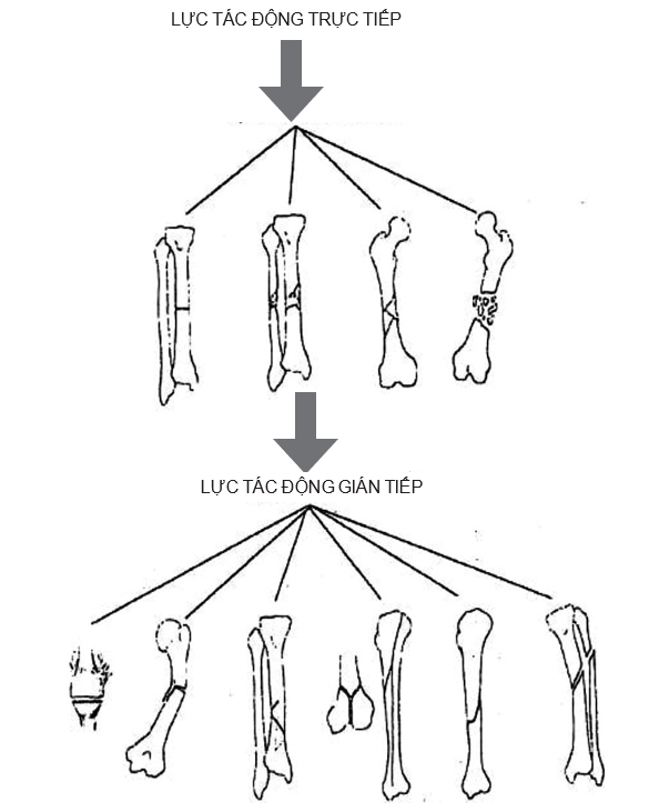
The vast majority of common fractures are traumatic fractures. The cause is an external force acting on normal healthy bone. The force of injury (called the traumatic agent) can produce:
- Direct fracture if the fracture site is at the exact spot where the traumatic agent was placed. For example: a car running over a thigh causes a femur fracture, falling on the heel to the ground causes a heel fracture.
- Indirect fracture if the fracture site is far from the point of placement of the traumatic agent. For example, fractures caused by a lever bending agent, fractures caused by twisting. Sometimes the cause of the fracture is the pulling force that causes the fracture. There are two types of pull-ups:
- The external traumatic agent causes the muscle to stretch and pull strongly, causing the bone to break where the tendon is attached. This is the case of a fracture of the humerus at the end of the triceps brachii.
- External trauma overstresses the ligament, and it is the strained ligament that pulls the bone at the end of the ligament. It is a case of chipping the bone where the lateral ligament 's baseball knee joint.
MECHANISM AND TYPES OF LINES
The causative agent of the fracture and the muscle response of the fractured limb form a fracture mechanism, as a general rule each type produces a typical fracture line:
- The mechanism that directly induces the bending action usually produces a transverse fracture (i.e., perpendicular to the longitudinal axis of the bony body).
- Indirect bending mechanism (lever type) often causes diagonal fractures
- Twisting mechanism, creating twisted fracture line
- The mechanism of squeezing and compressing can cause fractures or subsidence of bones
- Both bending, twisting and compression will cause a torsion fracture with a third wedge-shaped fracture (Figure 3.1).

Figure 3.1. Mechanism of injury related to closed fracture line type (Source: Fracture outline – Surgical Pathology – University of Medicine and Pharmacy, Ho Chi Minh City)
EFFECT OF GENDER AND AGE ON TYPE OF BACKGROUND
In general, both men and women and all ages experience traumatic fractures equally. But because the development of the skeleton has some age differences, there are some specific types of fractures.
In children
In children, the skeleton is growing, the periosteum is thickened, so the following types of fractures in the body can be encountered:
- Fracture en bois vert
- Traumatic bowing, fracture plastique.
At the ends of the bones, there is synaptic cartilage, so it is only in children that this type of "synaptic cartilage detachment" (see article Bone fractures in children).
In the elderly
In the elderly, there is a state of osteoporosis, so some weak cancellous bones are susceptible to fracture even with very minor trauma:
- Spondylolisthesis (back bend in the elderly)
- Fracture of femoral neck, humerus surgery neck, fracture of the lower radial head,...
In female
In women, after menopause, osteoporosis occurs earlier (compared to men of the same age), so fractures are more common.
TYPES OF BACKGROUND
Incomplete closed fractures (stem fractures mostly in children)
- Shaping fractures
- Hard cortical aneurysm fracture
- Fresh branches.
Completely broken
- Simple fracture (two segments)
- Two-stage fracture
- Multipart fracture (with a third piece, broken).
Special types of fractures
- Fracture with clasp
- Depression fractures
- Compression fracture
- Fracture of the synaptic cartilage in children.
TYPES OF DIFFERENT STRATEGY OF CLOSE BACKGROUND
Broken bones can stay in the same position, we call it a fracture without displacement. But many cases of fracture will be moved, we call fracture with displacement. The following five displacements can be distinguished:
Move to the side
The fracture is displaced perpendicular to the longitudinal axis of the bone.
Short axial displacement
Displaced fractures along the bone axis come close to each other. Called husband displacement for short.
Axial displacement away from each other
The fractures are displaced axially away from each other. Called distant displacement.
Angular displacement
The axis of the two fractures forms an angle (usually the acute angle).
Rotation displacement
Displaced distal fracture rotates around the longitudinal axis of the bone. A fracture may have one or more displacements (at most 4). When describing displacement, the convention says displacement of the distal fracture with respect to the proximal fracture.
IMPACTS OF BRAKES ON THE IMPORTED AREA AND THE WHOLE BODY
When there is a fracture, if only based on the film X-ray assessing bone damage would be a serious mistake. The external force that breaks the bone as well as the displacement of the broken bone segments creating additional internal trauma will affect all other tissues around the fracture site to some extent.
Effects on blood vessels
In any fracture, the blood vessels in the bone marrow, in the bone, and in the periosteum are broken. In addition, the force of trauma can cause additional bleeding in the surrounding soft tissues. Such bleeding forms a hematoma at the fracture site (called a fracture). If large bones are broken, heavy bleeding will cause significant blood loss and the victim can be stunned, (closed fracture does not see blood out, but the amount of hematoma in the fracture is no longer involved in circulation, so it is considered lost).
The table below shows a statistic by H. Willenegger on the extent of bleeding in some major fractures.
Table 3.1. Statistics of H. Willenegger on the degree of bleeding in some major fractures
|
Number of patients |
Amount of blood lost (mL) |
||||
|
Type of broken bone |
As soon as a bone breaks |
Three days later |
|||
|
Medium |
Max |
Medium |
Max |
||
|
Leg |
34 |
300 |
600 |
600 |
1.400 |
|
Femoral |
13 |
300 |
1.000 |
1.400 |
2.400 |
|
Pelvic |
13 |
1.700 |
2.400 |
2.500 |
4.000 |
Fractures with both broken blood vessels in the canal and contusion of the marrow, so it is possible that the fat of the bone marrow spills into the blood, causing fat vascular occlusion syndrome. Fat vascular occlusion syndrome due to fracture accounts for nearly 50% of total fat vascular occlusions is another possible cause of death in fracture victims.
If there is little hematoma in the fracture area, a few days after the injury, it will spread under the skin, creating signs of late bruising under the skin and then disappearing. If the hematoma is large, there will be swelling and bruising under the skin soon, creating pressure and obstructing local blood circulation. Excessive swelling can threaten to cause syndrome cavity compression heavier will cause caseation extremities where fractures occur, and may contribute to the nutritional disorder syndrome.
Compression of blood circulation due to hematoma also contributes to hypoxia in the fracture area, if significant deficiency will prevent the body from fighting infection if it is open fracture (see also open fracture article). If the main blood vessels are ruptured or perforated, the risks mentioned above are greater (sometimes the large vessels are only compressed by displaced fractures, early correction of displacements is the best way to avoid serious complications). ).
Impact on surrounding muscles
The muscles around the fracture area can be injured by the traumatic agent. In addition, the edema that compresses blood circulation can cause ischemia in the muscle and cause muscle necrosis or contracture. If the broken bone has displaced overlap, making the bone shorter, the surrounding muscles will relax and gradually shorten themselves. Any painful stimulus (broken bone cannot be motionless,…) càng làm cho các cơ co ngắn thêm. Như vậy, sự co cơ phản ứng này sẽ gây khó khăn cho điều trị kéo nắn các di lệch; để càng muộn sự co cơ càng nhiều thì kéo nắn càng khó (tốt nhất là nên kéo nắn cấp cứu sớm các gãy xương có di lệch gập góc và di lệch chồng ngắn, nhất là ở nạn nhân có các cơ to khỏe, khi các cơ chưa có phản ứng hoặc sự co cơ chưa mạnh).
Affects surrounding nerves
Nerves around the fracture can be damaged by direct or indirect causes of the fracture. Nerve damage can be torn, torn or overstretched.
Ischemic insertion can also cause neurological disorders. In this case, if the nerve is quickly released from the compression, permanent damage can be avoided.
Effects on the skin
If the fracture is accompanied by damage to the skin caused by external trauma or punctured by fractured segments, it is important to determine whether the skin is still covering the broken bone or if the skin damage has caused the fracture. fractures communicate with the outside. By convention, only when a skin wound causes the fracture to communicate with the outside, it is called an open fracture and only then does the fracture pose a threat of infection. An open fracture infection is a surgical infection.
In summary, complications of fractures are divided into two groups:
- Complications that immediately threaten the victim's life, include:
- + Injury shock
- Fat vascular occlusion syndrome.
- Complications that primarily affect the injured limb include:
- + Compulsion syndrome
- Injury to major major blood vessels
- Peripheral nerve damage
- + Open fractures and infections
- Nutritional disorder syndrome.
CLASSIFICATION OF BACKGROUND
The classification of fractures is based on several criteria, each with its own meaning. Thus a particular fracture can be classified in several ways. Here are some criteria for classification:
Clinically, the degree of soft tissue damage
Clinically, there are two basic types of fractures:
- Closed fracture: fracture without a skin wound or wound but not through the fracture (hematoma)
- Open fracture: A fracture has a skin wound and this wound penetrates the fracture site.
Dựa vào mức độ tổn thương mô mềm mà người ta chia gãy kín và broken open into many types.
Fracture grading according to OESTERNE and TECHERNE (1982): including four degrees
Table 3.2. Classification of fractures according to Oestem and Tscherne
|
Classification of fractures |
Soft tissue injury |
Bone damage |
Severity |
Common dangers |
|
|
Closed fracture |
Degree 0 |
Trivial |
Less displacement fractures |
+ |
Do not have |
|
Team |
Light touch |
Simple |
+ to ++ |
Few |
|
|
Degree II |
Medium touch |
Medium |
+ to +++ |
Cavity compression + |
|
|
Grade III |
Severely injured |
Complicated |
+ to +++ |
Cavity compression ++ |
|
Note: Severity: (+) light (++ ) moderate (+++ ) severe
According to the fracture location on the bone
- Fractures at the ends of bones:
- Extra-articular fracture
- Joint fracture.
- Fracture in the body of the bone:
- 1/3 above
- middle 1/3
- Lower 1/3.
The most commonly used classification is the AO (Swiss Association of Osteoarthritis) classification.
According to anatomical region
Depending on the fracture area has its own anatomical features, many study authors have produced their own classifications that are significant in terms of treatment and prognosis. Most of these classifications have their own names (usually the name of the person who first described the classification). For example, femoral neck fractures can be classified by Garden (four types) or by Pauwels (three types).
Based on the possibility of secondary displacement of closed fractures
It is also divided:
- Stable fractures: fractures are less likely to displace secondary to treatment
- Unstable fractures: fractures are more likely to displace secondary to treatment.
CLINICAL SIGNS OF CLOSE BACKGROUND
Sure sign
After an injury, if you see one or more of the following signs:
- Distortion (5 styles)
- Abnormal movements
- Bone crunching sound
Uncertain sign
- Painful
- Swollen, bruised
- Loss of ability.
TREATMENT
- Prediction of complications and prevention.
- Early implementation:
- Anesthetize the fracture with novocaine to relieve pain and relieve pain
- Good immobilization of the fracture area.


bài viết rất hay ạ
cảm ơn thầy ạ
Em cảm ơn ạ
Em cảm ơn tác giả ạ
Bài giảng rất hay ạ
cảm ơn thầy ạ
Bài giảng rất hay ạ
Cảm ơn thầy nhiều ạ
bài giảng rất bổ ích ạ
bài viết hay lắm ạ
bài viết rất hay ạ
hay thầy ạ
Bài giảng hay lắm ạ
Bài giảng rất hay ạ
Bài giảng rất hay ạ
cảm ơn thầy