Table of contents [hide]
SMALL ADMINISTRATIVE PROCEDURES
- Patient's name
- Patient number
- Date of shooting
- Number of movies
- Movie scale
- Movie on the right or on the left
- Film pose: normally there are two poses:
- + Straight shot: is shooting from front to back or vice versa.
- + Tilt: is shooting in the direction perpendicular to the film taken straight.
- Depending on the doctor's request, there are other shooting positions such as: cross-section, tangential, dynamic.
- Requirements of photographic film: a good film must meet three things:
- + Medium beam quality, not too old or not too young
- + Number of films: must have two straight and inclined films
- + Film size: must be long enough to be taken above one joint and below one joint taken from the injury site; wide enough to get the soft ball (in order to determine the position to avoid missing the lesion).
READ DAMAGE
Careful observation of all limb areas captured on radiographs including bones, joints, and soft tissues must be observed. In order to determine whether a lesion is present, it is necessary to know the normalization criteria for imaging on the respective limb regions of the normal person. Sometimes it is necessary to have a movie of ordinary people to compare and contrast. In children, it is a good practice to photograph both limbs in the same position.
If it's a broken bone
It is necessary to determine:
- Which bone is broken?
- Broken line
- Displacement degree.
Which bone is broken?
State the correct anatomical name of the bone.
Break location
According to the anatomical position. Long bones are divided into three parts as follows:
- Proximal head
- Bone body
- Distant head.
Distinguish the bone head from the body by the cortical bone. The bone body has thick cortical bone and no marrow canal, in the bone body people are divided into three regions: the upper 1/3, the middle 1/3 and the lower 1/3.
Thus, for a large long bone such as the femur, the fracture sites are as follows:
- Upper head
- Connecting the upper end to the upper 1/3
- 1/3 above
- Junction of upper 1/3 with middle 1/3
- middle 1/3
- Joint between the middle 1/3 and the bottom 1/3
- lower 1/3
- Joint of lower 1/3 with lower end
- Lower head.
The fracture site is also referred to by the specific anatomical name for that area. For example: femoral neck, femoral head, intertropical, below trochanter, above two condyle, medial condyle, lateral condyle, etc.
Broken line
The following types of fractures may be encountered:
- Incomplete fracture type:
- Fracture
- Broken
- Subsidence fracture
- Fracture of cortical aneurysm
- Formative fracture (no fracture line visible, only more curved bone)
- Fracture on one side of the shell.
- Completely broken type
- Transverse fracture: fracture line perpendicular to the longitudinal axis of the bone
- Cross fracture: fracture line is not perpendicular to the longitudinal axis of the bone
- Torsion fracture: spiral fracture line
- Fracture with 3rd fragment (butterfly): large fracture affects the stability of the bone
- Multi-fragmentation (fracture): large fractures that affect the stability of the bone
- Longitudinal fracture: fracture along the longitudinal axis of the bone
- Two-stage fracture: there are two fractures separated by a bony canal. If the bone is split vertically, it is a multi-fragment fracture
At the end of the bone, there can be many fracture lines, such as T, V, and Y fractures. Pay attention to whether the fracture line breaks the joint or not.
Based on the broken line to evaluate fracture stable or unstable.
Displacement
A broken bone may or may not be displaced. Incomplete fractures are usually nondisplaced, with the exception of fresh branch fractures with angular displacement. Complete fractures usually have little or no displacement. When reading displacement, attention should be paid to:
- Displacement type
- Displacement degree.
By convention, displacement orientation is based on displacement of the distal fracture with the proximal fracture. Fractures in the bony shaft can experience five types of displacement:
Move to the side
Displacement of the distal fracture inward, anteroposteriorly, relative to the proximal fracture. On straight film only the displacement is inward or outward. On slant film, only anterior or posterior displacement is determined.
Degree of displacement: take the width of the bone body as a standard to measure. We have levels: one cortical, 1/2 cortical, 1/4 bony, 1/3 bony, 1/2 bony, 2/3 bony, 1 bony, over 1 bony. The degree of displacement indicates the contact of the two ends of the broken bone to assess the ability bone healing and be sure. The length unit is not used to measure lateral displacement because the same displacement on a large bone can be a small displacement, but on a small bone a large displacement.
Displaced husband
Also called axial displacement near each other. The degree of displacement in mm, must determine the position to be measured based on the fracture line. In case of multi-fragmentation fracture, the degree of displacement cannot be determined (only determined by clinical limb measurement).
Angular displacement
The limb axis of the distal segment is not on the limb axis of the proximal fracture but forms an angle.
- Displacement direction: is the direction of the angle with the limb axis of the two fractures.
- Displacement degree: is the measured value of the angle between the axis of the distal fracture and the extension of the axis of the proximal fracture.
Rotation displacement
Is the displacement of the distal fracture about its longitudinal axis. It is only necessary to determine whether there is rotational displacement or not, but the degree of displacement is difficult to measure accurately. To determine rotational displacement, it is necessary to prove that the two fractures are not in the same plane on the same radiograph. This can be seen:
- Two fractures are not in the same plane if two joints are taken at both ends
- There is a clear difference in the diameter of the bone body near the fracture line of the two segments
- The two-segment broken line doesn't match
- In the fracture area with two bones of the forearm or lower leg, the parallel passage of the two bones is not the same in the two fractures.
Displacement
Còn gọi là di lệch dọc trục xa nhau thường do cơ co kéo (gãy xương bánh chè, mỏm khuỷu, củ lớn heel bone,...) or by muscle insertion into the fracture. This displacement has the risk of causing broken bones to not heal because the two ends of the broken bones are not in contact. Degree of displacement in mm.
A fracture can have one to four displacement patterns because superimposed displacement has no distal displacement (not to be confused with lateral displacement above a bony body as distal displacement).
The penetrating fractures must be examined for articular facet canal leveling.
For two-stage (or multi-layered) fractures, the displacement of each fracture should be read and then summed up as the displacement of the farthest fracture for the nearest fracture. The degree of displacement added includes: displacement, overlapping, and rotation. Lateral and distal displacements do not add up because these displacements are significant for each fracture (evaluate the healing ability at each fracture).
The image printed on the film is a projection of all the parts in the imaging area that the X-rays pass through and affect the film. On conventional films (no zooming in or out) the imaged organs are usually slightly larger than the actual size of the organs, depending on the position of the lamp head, the position of the film, and the organ being imaged (because the beam is emitted in the form of a beam). divergence). For an approximate size, ask the photographer to take a distance shot. If the physician uses a ruler to measure the size of the bone or joint to select the surgical instrument in advance, it should subtract about 10% from the measured size.
Read Structural Abnormalities
Common in pathological fractures or old dislocation fractures with complications. Changes in bone structure can be seen on images X-ray based on the change in its radiopaque properties, including:
Bone resorption
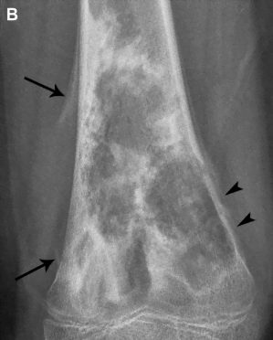
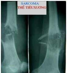
Figure 13.1. Bone graft surgery [2]
The loss of the entire structure of an area of bone, the bone resorption can be in the head or body, in the bone marrow or in the cortex. Boundaries are often jagged or sharp (as in the resorption foci of Kahler disease). In bone resorption, the contrast density can be uniform or septum, sometimes there is calcification or dark contrast nodule formed by dead bone fragments. Bone resorption is common in primary or secondary osteolytic malignancies.
Bone thickening (due to osteogenic reaction)
Bone thickening can occur starting from the trabecular or from the medial surface of the periosteum. Bone thickening is common in alveolar fractures, osteomyelitis in the chronic stage, and primary or metastatic osteosarcoma. If bone thickening occurs in the long canal, it may obscure the canal.
Figure 13.2. Thick bone [2]
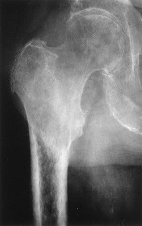
Osteoporosis
It is a phenomenon of decreased bone calcium, common in osteoporosis in the elderly, due to motionless long-term fracture, early stage of tuberculosis joints and bone marrow inflammation, etc. Due to the decrease in calcium density of bone, bone ridges and trabecular bones are often clearly visible on radiographs.
Figure 13.3. Osteoporosis [2]
Dead bones
A condition in which the bone structure is only present with minerals and no organic matter. Dead bone may appear in the inflamed bone marrow caseation aseptic cartilaginous end ossification and the ossicles are in the process of ossification.
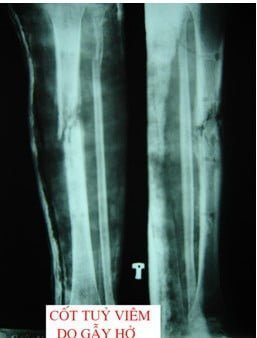
Figure 13.4. Dead Bones [2]




The post is useful