Table of contents [hide]
ACCESS TO DIAGNOSIS
- Main symptom: pain shoulder
- Common diseases: arthritis vai, hẹp mỏm cùng vai, tổn thương SLAP, viêm gân nhị đầu
- Uncommon disease: sacroiliitis
- Patient identification: middle age, shoulder pain, no sign of numbness in hands
PATHOLOGICAL
Outline
1. Definition:
- A rotator cuff tear is a tear in one or more tendons in the rotator cuff tendon complex.
-
The rotator cuff is a complex at the shoulder joint consisting of four muscle tendons: the supraspinatus, subspinous, small round, and subscapular tendons. This structure contributes an important part to the complex functioning of the shoulder joint. Injury to the rotator cuff is one of the most common features of the shoulder joint, resulting in a reduction or complete loss of range of motion. In which, the main injury is tearing at the attachment point of the tendon.
2. Damage mechanism
- Studies around the world show that partial or complete rotator cuff tear is mainly caused by three main factors: trauma, endogenous, exogenous
- Endogenous factors related to blood supply problems or metabolic problems that develop with age, leading to degenerative tears
- The extrinsic factor is usually due to narrowing of the apical space of the shoulder, causing the rotator cuff to be irritated and rubbed continuously, leading to tearing
- Injury factors are strong impacts that cause acute damage to the rotator cuff tendon.
Pathophysiology of rotator cuff tear
- The rotator cuff tendon has a mucilaginous and elastic structure that can withstand very high tension up to 100 N/mm. However, the ability to withstand compressive and tearing forces is less.
- In cadaver shoulder studies, maximum tension in the rotator cuff tendons was achieved when the arm was at 120 flex.o.
- Degeneration accompanied by extrinsic compression or eccentric overload will cause the rotator cuff tendon to tear
Classification of rotator cuff tear
There are many ways to classify rotator cuff tears depending on the concept of each author. In practice, it is often based on the thickness and location of the tear, size, and shape
1. According to thickness and position
- Partial tear at the joint face
- Partial tear on the underside of the apex of the shoulder
- Tear in tendons
- Totally torn
2. According to size
- Small tear: < 1 cm
- Medium tear: 1-3 cm
- Large tear: 3-5 cm
- Very large tear: > 5 cm.
3. By shape: sickle, U, L, very big tear
ASSESSMENT OF PATIENTS
Medical history: Ask for a history of shoulder injury. If there is a history of trauma, we will learn more about the time, mechanism, and what has been treated after the injury.
Clinical examination: should pay attention to the following examination tests:
- Comparative external rotation test: patient is shoulder flexed, elbow flexed 90o, rotate out. The physician will resist the patient's external rotation. If the patient is in pain, a positive test indicates that the patient is likely to tear the apex
- Jobe's test: the patient is 90° outstretchedo. The doctor will press the patient's hand down, the patient resists the pressure. If the patient is in pain, a positive test indicates that the patient is likely to tear the apex
- Falling arm test: the doctor lifts the patient's arm up and then releases it. If the patient is unable to hold the arm in this position and falls down, it will be positive. Thus, there is a possibility that the rotator cuff will be torn.
Subclinical
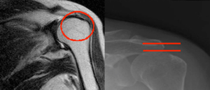
- X-ray Shoulder joint three positions: straight, inclined,
- Magnetic resonance (MRI): has the highest accuracy. On MRI, it will show the tendon tear morphology, the degree of tendon tear, the number of torn tendons, the associated injuries

DIAGNOSE
Differential diagnosis
Lesions of SLAP; biceps tendonitis; narrow at the shoulder.
Implementing the quadrants
- Shoulder pain, positive physical exam
- MRI tear apical tendon
Diagnose the cause: due to trauma or degeneration
TREATMENT
Purpose: to help patients recover from illness
Rule: start medical treatment then go to surgical treatment
Specific treatment
1. Conservative treatment: anti-inflammatory, analgesic, physiotherapy
- Case of partial tear of the rotator cuff tendon, rotator cuff tendinitis
- Severe medical disease that cannot be surgically intervened.
2. Surgical treatment: laparoscopic surgery, or open surgery
- In case of complete tear of the rotator cuff tendon
- Partial tear of the rotator cuff tendon but failed medical treatment (medical treatment duration > 03 months).
Currently, laparoscopic surgery is widely applied in hospitals around the world as well as in Vietnam because of the following advantages:
- Minimally invasive surgery
- High aesthetics
- Short hospital stay
- Monitor and recover all damage.
How to perform laparoscopic surgery:
- Preparing the patient for surgery
- Prepare complete machinery and equipment
- Endotracheal anesthesia patient
- Enter the joint through the endoscopic routes, to investigate the damage
- Getting the anchor thread to the position of the tendon attachment point
- Sewing torn rotator cuff tendons
- Hemostasis, skin suturing, sterile dressings.
FOLLOWING RE-examination and rehabilitation exercise
– Exercise to maintain joint range: 4-6 weeks
+ 1st week:
- Wear splint
- Shrug your shoulders, strain your muscles
- Tập chủ động ngón tay, khuỷu tay, cổ
+ Weeks 2 - 3:
- Wear splint
- Shrug your shoulders, strain your muscles
- Active exercise fingers, elbows, wrists → Practice hand walking on the wall, pull the pulley.
+ Week 4 - 5:
- Remove splint
- Practice hand walking on the wall, pull the pulley
- Maximum restored joint range of motion.
- Strength training: from week 6 onwards
+ Maintain joint range of motion
+ Strength training with elastic bands
+ Strength training with weights.


